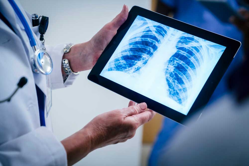
X-ray Chest
A Chest X-ray is a quick and essential imaging test used to examine the lungs, heart, and surrounding structures. It helps detect infections, lung diseases, fractures, fluid accumulation, and other abnormalities that may affect your breathing or overall chest health. With its ability to provide detailed internal images in just a few seconds, a Chest X-ray plays a vital role in diagnosing conditions like pneumonia, tuberculosis, heart enlargement, and rib injuries. It is a safe, noninvasive, and reliable test that supports accurate medical evaluation and timely treatment.
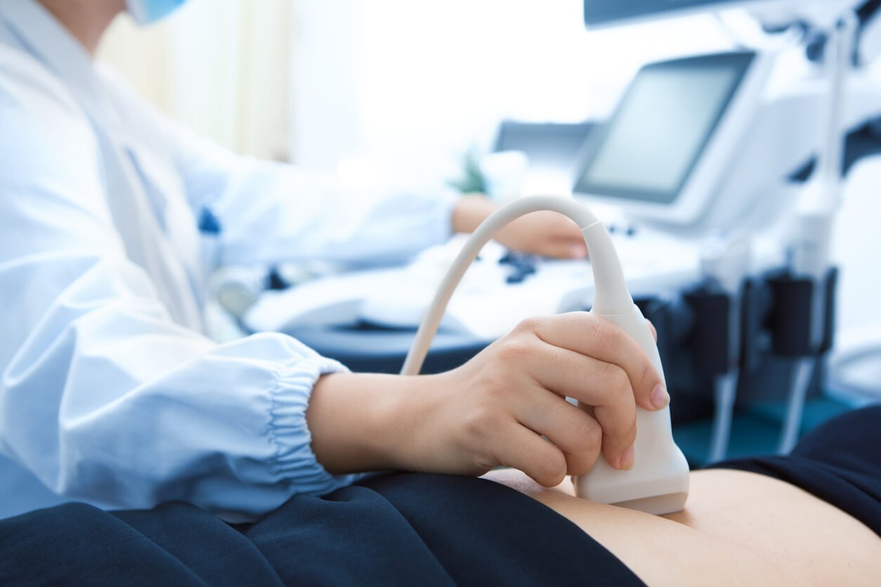
Sonography
Sonography is a safe, painless imaging technique that uses sound waves to capture detailed pictures of your internal organs. It helps doctors evaluate the abdomen, pelvis, soft tissues, pregnancy, and many other areas without exposing you to radiation. Sonography is commonly used to detect abnormalities, monitor ongoing conditions, and guide precise medical decisions. With quick results and high accuracy, it is one of the most reliable diagnostic tools for early detection and effective treatment planning.
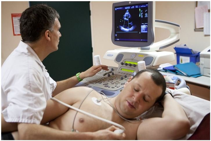
2DEcho Cardio
2D Echo, or Echocardiography, is a specialized ultrasound test that provides real-time images of your heart. It helps assess the structure and function of the heart chambers, valves, and blood flow. This painless, noninvasive test is essential for detecting conditions like valve disorders, heart muscle weakness, congenital defects, and fluid around the heart. With detailed visual insights, a 2D Echo allows doctors to make accurate diagnoses and plan effective treatment, ensuring better long-term heart health. It helps evaluate how well treatments are working, track changes in heart function, and identify potential problems before they become serious. Regular echocardiography is especially beneficial for individuals with hypertension, diabetes, previous heart disease, or symptoms like breathlessness and fatigue.